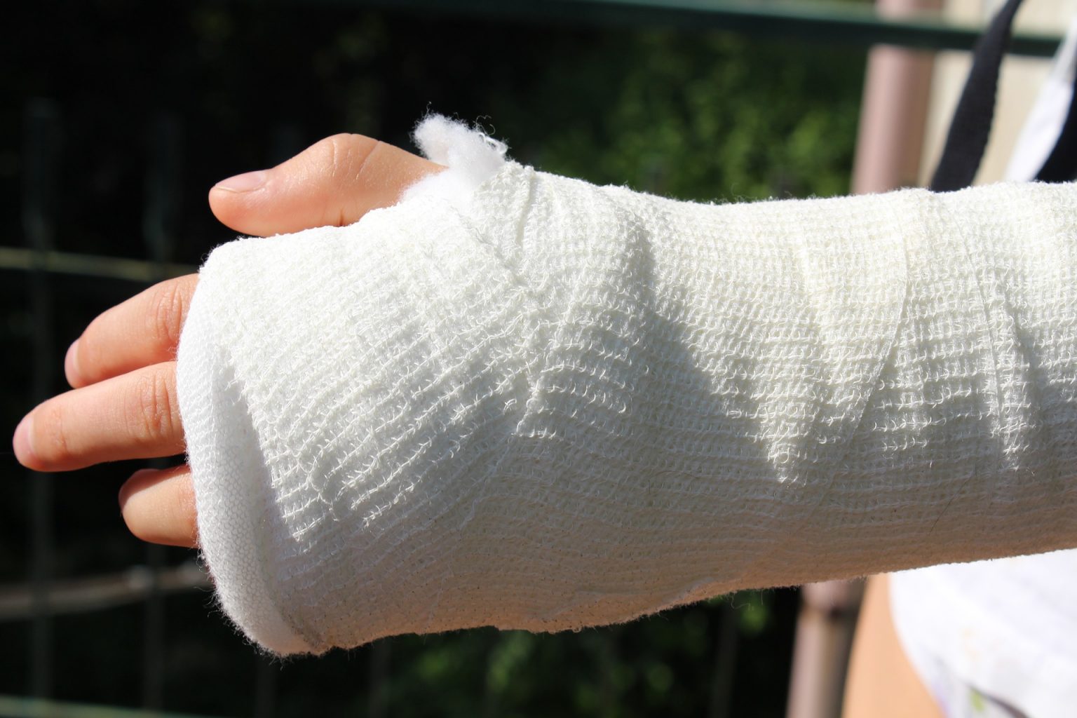
Severe dysfunctions may be caused by proximal forearm fractures. These dysfunctions are caused by post-traumatic instability, mal-union, or non-union and impingement. Different articulations in the elbow constitution may be involved in these injuries such as: the ulnotrochlear joint, the proximal radioulnar joint or the radiocapitellar joint. The structural anatomy of the elbow joint should be considered carefully.
The common fractures are osteochondral or loose bodies, particularly with radial head injuries related to capitellar abrasions. Distal injuries like forearm fractures, distal radius, ruptures of the interosseous membrane, and distal radioulnar joint disruption can be the result of higher energy fractures. These injuries should be diagnosed with the help of the x-ray of the entire forearm. Surgeon use Microlock locking hand plate for the hand fracture surgery.
It is very important to attain adequate anatomical reconstruction of the different ring structures to promote early functional treatment which needs both reduction of the osseous components and the restoration of the tension of the ligamentous and capsular avulsions.
Sometimes, badly comminuted fragments can be excised in the absence of associated ligamentous instability. A comminuted olecranon in elderly patient can be treated by resection and reattachment of the triceps, if no fractures of the coronoid process or avulsion of the anterior capsule exist. And if the comminuted radial head is not manageable for reconstruction, a prosthetic spacer can be used to replace it. This will facilitate the healing of the torn capsule or ligaments without compromising the function of the elbow or forearm.
Classification of proximal forearm fractures can be explained as vulnerable to direct trauma, torsions and hyperextensions. The most common of elbow injuries are fractures of the olecranon.
Assessment of fractures and soft tissues
Generally, the fracture indicates disruption of the triceps mechanism along with a bending moment over the distal end of the trochlea which induce the oblique B1 patterns and characteristic transverse. Comminution and impaction of the central part of the olecranon articular surface is generated by the more direct forces and sometimes avulsions of the coronoid process also.
The patient finds himself unable to use the elbow due to severe pain. There may be The swelling, contusion, or bruising onto the skin. The lateral view can show the fracture line, the amount of displacement clearly and the degree of comminution if any. A lateral tomogram proves very helpful in the assessment of the degree of articular impaction. An anterior dislocation of the forearm or a posterior type II Monteggia lesion may be associated with complex fractures.
Despite of so many classification of fractures, one cannot count on simple transverse or oblique fractures to represent the typical stable pattern because these fractures can be associated with elbow or forearm dislocations. Similarly, more stability can be seen in multi-fragment fractures if confined to the trochlear notch and if the coronoid process or proximal ulna are involved, they may be grossly unstable.
Preoperative planning:
Positioning and approaches
As far as the positioning of the patient is concerned, the patient must be in the lateral position with the elbow flexed over a side rest or he must be in prone position. The other option is the supine position with the forearm placed across the chest particularly with extended approaches to the lateral pillar or column. After preparing the skin and draping, a sterilized tourniquet is placed on the upper arm. The skin incision runs from the supracondylar area posteriorly to a point 4 or 5 cm distal to the fracture. In order to protect the ulnar nerve and to avoid skin bruises or lacerations, it can be curved gently to the radial side. Large skin flaps should be avoided as they do not heal finely.
Reduction tools and techniques
Generally, indirect reduction technique is used for multi-fragmentary fractures. While for articular fractures, direct reduction technique using hooks, K-wires and pointed reduction forceps, is used.
Radial head prosthesis implants are useful for severe elbow traumatic instability and failure of osteosynthesis. Locking proximal medial tibia plates are indicated for metaphyseal fractures of the medial tibial plateau, fixation of the proximal lateral and medial of the tibia and non-unions and mal-unions of the medial proximal tibia.

